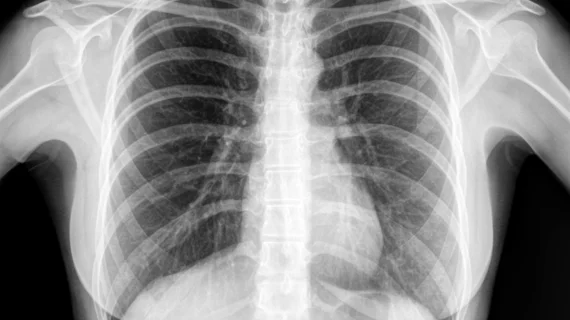
AI models can be trained to evaluate chest x-rays as well as radiologists, according to a new study published by Radiology. Specialist-approved reference standards played a crucial role in the team’s research.
The study’s authors—including several researchers from Google Health in Palo Alto, California—worked to overcome certain restrictions commonly associated with how radiologists interpret chest x-rays.
“We've found that there is a lot of subjectivity in chest x-ray interpretation,” co-author Shravya Shetty, an engineering lead for Google Health, said in a prepared statement. “Significant inter-reader variability and suboptimal sensitivity for the detection of important clinical findings can limit its effectiveness.”
Two massive datasets were used for the research, one that included 750,000 images from India and another that included more than 100,000 images from the National Institutes of Health (NIH). A team of radiologists then worked together to develop reference standards related to four findings—pneumothorax, opacity, nodule or mass, and fracture—commonly found on chest x-rays.
The radiologist input made a significant impact, helping the researchers achieve an overall consensus of 97%. The team also noted that the AI models detected findings missed by radiologists and their radiologists caught things missed by the models, “suggesting that machine learning–based assistive tools could potentially improve performance over either models or radiologists alone.”
“We believe the data sampling used in this work helps to more accurately represent the incidence for these conditions,” Daniel Tse, MD, product manager for Google Health, said in the same statement. “Moving forward, deep learning can provide a useful resource to facilitate the continued development of clinically useful AI models for chest radiography.”
“We hope that the release of our labels will help further research in this field,” Shetty concluded.

