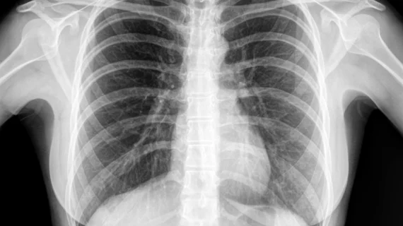
AI-powered software can help radiologists identify malignant lung cancers with improved accuracy, according to new research published in Radiology.
The study’s authors explored data from four different hospitals, including 600 randomly selected chest x-rays featuring at least one malignant lung nodule and another 200 normal chest x-rays. All exams occurred between October 2015 and September 2017.
A group of radiologists then read each study twice—once on their own and once with assistance from a deep convolutional neural network (DCNN) trained specifically to detect lung nodules. The DCNN was trained on more than 13,000 normal x-rays and another 3,500 x-rays containing lung nodules.
Overall, the radiologists saw their average false-positive findings drop from 0.2 per x-ray to 0.18 per x-ray and average sensitivity jump from 65.1% to 70.3%.
“Computer-aided detection software to detect lung nodules has not been widely accepted and utilized because of high false positive rates, even though it provides relatively high sensitivity,” co-author Byoung Wook Choi, MD, PhD, department of radiology at the Yonsei University Health System in Seoul, Korea, said in a prepared statement. “DCNN may be a solution to reduce the number of false positives.”
The authors did note that their research had several limitations. For example, their dataset was “prone to spectrum bias” because certain images showing benign nodules were removed before the study began. In addition, no specific time was established that had to occur between the radiologists’ two interpretations, “leading to the possibility of recall bias.”

